Showing 31–59 of 59 results

Atlas of Anatomy of the Peripheral Nerves: The Nerves of the Limbs – Student Edition

Price range: ₨ 400 through ₨ 900
Black & White Print
Color Matt Paper Print
Clear
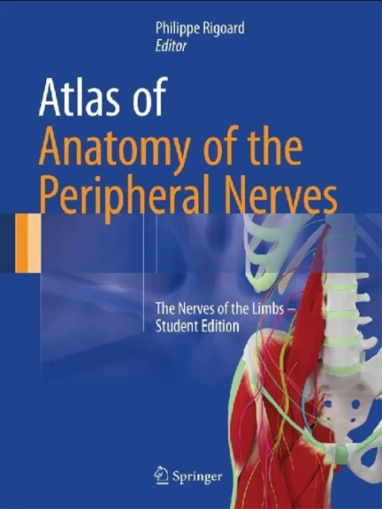
Atlas of Anatomy of the Peripheral Nerves: The Nerves of the Limbs – Student Editionby Philippe Rigoard (Editor) the booklanceThis innovative atlas focuses on peripheral nerves and provides a brand new approach compared to regular anatomy books. Using a modern 3D approach, it offers an alternative to conventional anatomical structures. It reviews all the anatomy and the morphology of these structures from an original point of view. In these three-dimensional diagrams, as well as in the watercolor drawings enhanced with a 3D inlay, each type of nerve is depicted in a minute detail. The atlas simplifies the anatomy and make it easy and understandable by allowing readers to develop a mental “real-time 3D GPS”. The integration of MRI sections related to the drawings and the descriptions of the main nerve injuries provide medical students with a flexible but effective transition to the radiological interpretation and furthers the clinical learning process. After a detailed evaluation of the morphofunctional anatomy of the peripheral nerves, the authors present a collection of relevant data on neuromuscular transmission, both from classical and recent literature, ranging from the central and peripheral nervous system to the effector muscle. This information offers a basis for understanding the physiology, the pathology, and the repair prospects of peripheral nerves from a purely theoretical point of view.The book is divided into three main parts: – Fundamental notions: from immunohistochemistry to limb innervation- The upper limb: the brachial plexus and related peripheral nerves- The lower limb: the lumbosacral plexus and related peripheral nervesThis atlas also features 261 outstanding full-colour 2D and 3D illustrations. Each picture has been designed in 2D and 3D with a combination of the original editor’s personal drawings/paintings and 3D-modeling tools. This book is a valuable resource for anyone studying medicine, anaesthesiology, neurosurgery, spine surgery, pain, radiology or rheumatology and is also of high interest to the whole medical community in general.

Atlas of Anatomy of the Peripheral Nerves: The Nerves of the Limbs – Student Edition

Price range: ₨ 600 through ₨ 1,200
Black & White Print
Color Matt Paper Print
Clear
Atlas of Anatomy of the Peripheral Nerves: The Nerves of the Limbs – Student Editionby Philippe Rigoard (Editor) the booklanceThis innovative atlas focuses on peripheral nerves and provides a brand new approach compared to regular anatomy books. Using a modern 3D approach, it offers an alternative to conventional anatomical structures. It reviews all the anatomy and the morphology of these structures from an original point of view. In these three-dimensional diagrams, as well as in the watercolor drawings enhanced with a 3D inlay, each type of nerve is depicted in a minute detail. The atlas simplifies the anatomy and make it easy and understandable by allowing readers to develop a mental “real-time 3D GPS”. The integration of MRI sections related to the drawings and the descriptions of the main nerve injuries provide medical students with a flexible but effective transition to the radiological interpretation and furthers the clinical learning process. After a detailed evaluation of the morphofunctional anatomy of the peripheral nerves, the authors present a collection of relevant data on neuromuscular transmission, both from classical and recent literature, ranging from the central and peripheral nervous system to the effector muscle. This information offers a basis for understanding the physiology, the pathology, and the repair prospects of peripheral nerves from a purely theoretical point of view.The book is divided into three main parts: – Fundamental notions: from immunohistochemistry to limb innervation- The upper limb: the brachial plexus and related peripheral nerves- The lower limb: the lumbosacral plexus and related peripheral nervesThis atlas also features 261 outstanding full-colour 2D and 3D illustrations. Each picture has been designed in 2D and 3D with a combination of the original editor’s personal drawings/paintings and 3D-modeling tools. This book is a valuable resource for anyone studying medicine, anaesthesiology, neurosurgery, spine surgery, pain, radiology or rheumatology and is also of high interest to the whole medical community in general.

Atlas of Botulinum Toxin Injection, Dosage, Localization, Application, 3rd Edition

Price range: ₨ 500 through ₨ 1,100
Black & White Print
Color Matt Paper Print
Clear
Atlas of Botulinum Toxin Injection, Dosage, Localization, Application, 3rd Editionby Wolfgang Jost (Author), Klaus-Peter Valerius (Contributor) the booklanceThis comprehensive atlas, now in its third edition, presents all of the information necessary for botulin toxin use, including dosage, muscle action, localization, and injection technique. Designed for quick navigation, it details the treatment of 126 different muscles organized by body region, illustrating muscle action, injection sites, injection protocols, and injection techniques for each muscle. Revisions to this new edition include updated and refined illustrations and expanded product information.Contents: 1. Introduction 2. Upper Limbs 3. Lower Limbs 4. Trunk 5. Neck 6. Larynx 7. Head 8. Pelvic Floor 9. Vegetative Indications Appendices

Atlas of Cardiac Anatomy: Anatomical Basis of Cardiac Interventions, Volume 1 1st Edition

Price range: ₨ 700 through ₨ 1,400
Black & White Print
Color Matt Paper Print
Clear
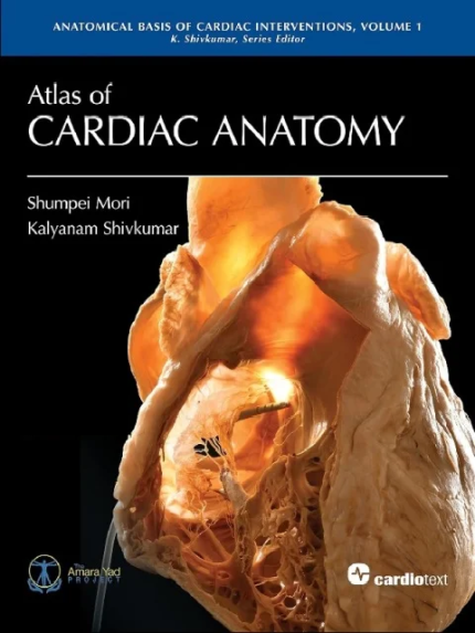
Atlas of Cardiac Anatomy: Anatomical Basis of Cardiac Interventions, Volume 1 1st Editionby Shumpei Mori (Author), Kalyanam Shivkumar (Author) the booklanceAtlas of Cardiac Anatomy represents a new standard for the study of the human heart.Drs. Shumpei Mori and Kalyanam Shivkumar have created a visually stunning presentation of more than 200 full-color photographs of the human heart and its adjacent structures.Atlas of Cardiac Anatomy includes unpublished and restored iconic photographs from the Wallace A. McAlpine Collection at UCLA and new images from the UCLA Cardiac Arrhythmia Center based on the methods and photographic techniques for the study of cardiac anatomy pioneered by Dr. McAlpine.This first volume serves as a core foundational study of cardiac anatomy and provides a basis for planned atlases edited by Dr. Shivkumar in the series, Anatomical Basis of Cardiac Interventions. Electrophysiology, structural heart disease, imaging, and cardiac surgery are the focus of future atlases.The authors’ goals are to offer an exceptionally comprehensive resource on cardiac anatomy. It aims to inspire new solutions and provide additional perspectives for skilled interventional and surgical operators. More broadly, it is likely to be useful for anyone caring for patients with cardiac disease.Includes access to 25 anaglyphs (3D images). 3D glasses required for viewing and are not included.The authors are at the UCLA Cardiac Arrhythmia Center, Los Angeles, California.Foreword by Francis E. Marchlinski, MD, and William G. Stevenson, MD.

Atlas of Endoscopic Neurosurgery of the Third Ventricle: Basic Principles for Ventricular Approaches and Essential Intraoperative Anatomy 1st eds

Price range: ₨ 500 through ₨ 1,000
Black & White Print
Color Matt Paper Print
Clear
Atlas of Endoscopic Neurosurgery of the Third Ventricle: Basic Principles for Ventricular Approaches and Essential Intraoperative Anatomy 1st edsby Roberto Alexandre Dezena (Author) the booklanceThis book describes in practical terms the endoscopic neurosurgery of the third ventricle and surrounding structures, emphasizing aspects of intraoperative endoscopic anatomy and ventricular approaches for main diseases, complemented by CT / MRI images. It is divided in two parts: Part I describes the evolution of the description of the ventricular system and traditional ventricular anatomy, besides the endoscopic neurosurgery evolution and current concepts, with images and schematic drawings, while Part II presents a collection of intraoperative images of endoscopic procedures, focusing in anatomy and main pathologies, complemented by schemes of the surgical approaches and CT / MRI images.The Atlas of Endoscopic Neurosurgery of the Third Ventricle offers a revealing guide to the subject, addressing the needs of medical students, neuroscientists, neurologists and especially neurosurgeons.

Atlas of Functional Shoulder Anatomy

Price range: ₨ 450 through ₨ 850
Black & White Print
Color Matt Paper Print
Clear
Atlas of Functional Shoulder Anatomy the booklanceby Giovanni Di Giacomo (Editor), Nicole Pouliart (Editor), Alberto Costantini (Editor), Andrea de Vita (Editor)The anatomy of the shoulder is based on complex joint biomechanics. The purpose of this Atlas is to focus the reader’s attention on a series of bone, ligament, muscle and tendon structures and ultrastructures within the shoulder on which only the most recent international literature has reported in specialized journals. This Atlas also presents extremely high-definition images of “targeted” sections obtained from cadavers preserved using state-of-art techniques. This unique Atlas, making use of images of major visual impact, offers a scientific message on a topical joint, using simple but dedicated descriptive language.

Atlas of Human Anatomy 1st Edition

Price range: ₨ 600 through ₨ 1,300
Black & White Print
Color Matt Paper Print
Clear
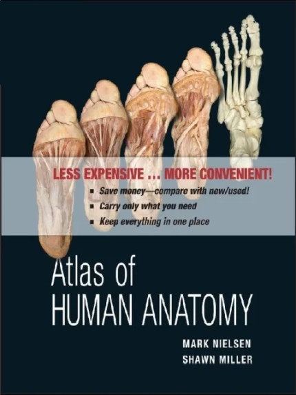
Atlas of Human Anatomy 1st Editionby Mark Nielsen (Author), Shawn D. Miller (Author) the booklanceThis book will motivate and engage health professionals to learn the essentials of anatomy and physiology through its visual approach and special pedagogy. The condensed content covers the basics without extensive detail. Core concepts are presented visually to enable them to gain a better understanding of the material. Process diagrams are integrated throughout the chapters to guide health professionals through the material.

Atlas of Imaging Anatomy 2015th Edition

Price range: ₨ 500 through ₨ 1,000
Black & White Print
Color Matt Paper Print
Clear
Atlas of Imaging Anatomy 2015th Editionby Lucio Olivetti (Editor) the booklanceThis book is designed to meet the needs of radiologists and radiographers by clearly depicting the anatomy that is generally visible on imaging studies. It presents the normal appearances on the most frequently used imaging techniques, including conventional radiology, ultrasound, computed tomography, and magnetic resonance imaging. Similarly, all relevant body regions are covered: brain, spine, head and neck, chest, mediastinum and heart, abdomen, gastrointestinal tract, liver, biliary tract, pancreas, urinary tract, and musculoskeletal system. The text accompanying the images describes the normal anatomy in a straightforward way and provides the medical information required in order to understand why we see what we see on diagnostic images. Helpful correlative anatomic illustrations in color have been created by a team of medical illustrators to further facilitate understanding.

Atlas of Lymphatic Anatomy in the Head, Neck, Chest and Limbs 1st ed

Price range: ₨ 500 through ₨ 1,000
Black & White Print
Color Matt Paper Print
Clear
Atlas of Lymphatic Anatomy in the Head, Neck, Chest and Limbs 1st edby Wei-Ren Pan (Author) the booklanceThis atlas provides detailed information on the human lymphatic system in the head, neck and chest regions as well as the extremities, with more than 400 photographs and radiographs, including micro and macro views of the morphology. Much of the content is presented for the first time, such as the individual differences in lymphatic distribution, especially in the head neck region; characteristics of the indirect precollecting lymph vessel in the scalp; the lymphatic ampulla and diverticulum; and the transparent lymph node. Providing insights into the lymphatic anatomy, the book is an essential resource for medical and science students as well as therapists, clinicians and researchers working in this field.

Atlas of Ocular Anatomy 1st ed. 2016 Edition

Price range: ₨ 250 through ₨ 400
Black & White Print
Color Matt Paper Print
Clear
Atlas of Ocular Anatomy 1st ed. 2016 Editionby Mohammad Wakeel Ansari (Author), Ahmed Nadeem (Author) the booklanceThis book is a practical and concise atlas on ocular anatomy, with an emphasis on applied aspects and hints for easy retention strategies. The vast color illustrations and photographs consist of self-explanatory, precise, and meaningful representations of the points covered in the text. Covering chapters such as bony socket of the eye, extraocular muscles, eyelids, cornea and lens, and neurology of the eye, Atlas of Ocular Anatomy gives a summary of the important and relevant points for each topic, separating out the essential from the nonessential elements. Complete with representative schematic line diagrams and full color photographs, this atlas features the correlation between anatomic facts with their probable clinical presentations in disease.

Atlas of Sectional Anatomy: Understanding the Anatomical Aspects of the Thorax, Abdomen and Pelvis 1st ed. 2022 Edition

Price range: ₨ 200 through ₨ 400
Black & White Print
Color Matt Paper Print
Clear
Atlas of Sectional Anatomy: Understanding the Anatomical Aspects of the Thorax, Abdomen and Pelvis 1st ed. 2022 Editionby Luciano Alves Favorito (Editor), Natasha T. Logsdon (Editor) the booklanceSectional anatomy is a valuable resource for understanding and interpreting imaging exams, specially computed tomography (CT) and magnetic resonance imaging (MRI). Thus, health professionals should have a solid anatomical knowledge to properly evaluate such exams during clinical assessments of cardiac, thoracic, abdominal, proctologic, gynecological and urological diseases.The chapters in this book describe the thoracic anatomy, the abdominal wall, retroperitoneal space, and the male and female pelvis. Sectional images of cadaveric material illustrate the thoracic and the abdominal cavities, kidney, ureter, prostate, penis and other male and female organs. The images and descriptions build familiarity with the anatomical traits and can be applied in the fields of urology, gynecology, proctology, radiology and surgery.This work appeals to a wide range of readers, from health professionals to residents and students of different medical specialties.

Atlas of Stem Anatomy in Herbs Shrubs and Trees Vol 1

Price range: ₨ 800 through ₨ 1,800
Black & White Print
Color Matt Paper Print
Clear
Atlas of Stem Anatomy in Herbs Shrubs and Trees Vol 1by Fritz Hans Schweingruber (Author), Annett Börner (Author), Ernst-Detlef Schulze (Author) the booklanceThis work, published in two volumes, contains descriptions of the wood and bark anatomies of 3000 dicotyledonous plants of 120 families, highlighting the anatomical and phylogenetic diversity of dicotyledonous plants of the Northern Hemisphere. The first volume principally treats families of the Early Angiosperms, Eudicots, Core Eudicots and Rosids, while the second concentrates on the Asterids.Presented in Volume 1 are microsections of the xylem and phloem of herbs, shrubs and trees of 1200 species and 85 families of various life forms of the temperate zone along altitudinal gradients from the lowland at the Mediterranean coast to the alpine zone in Western Europe. The global perspective of the findings is underlined by the analysis of 500 species from the Caucasus, the Rocky Mountains and Andes, the subtropical zone on the Canary Islands, the arid zones in the Sahara, in Eurasia, Arabia and Southwest North America, and the boreal and arctic zones in Eurasia and Canada.The presence of annual rings in all life forms demonstrates that herbs and dwarf shrubs are an excellent tool for the reconstruction of annual biomass production and the interannual dynamic of plant associations. The common principle of the anatomical expression of secondary growth is a key factor in understanding evolution and adaptation processes in all life forms, from the 2 cm tall whitlow grass (Draba arctica) in the arctic to the 40 m tall beech (Fagus sylvatica) in Central European managed forests. The study opens vast fields of research for dendrochronology, wood anatomy, taxonomy and ecology.

Atlas of the Human Body: Central Nervous System and Vascularization 1st Edition

Price range: ₨ 500 through ₨ 1,000
Black & White Print
Color Matt Paper Print
Clear
Atlas of the Human Body: Central Nervous System and Vascularization 1st Editionby Branislav Vidic (Author), Milan Milisavljevic M.D. S.D. D.Sc. (Author) the booklanceAtlas of Human Body: Central Nervous System and Vascularization is a multidisciplinary approach to the technical coverage of anatomical structures and relationships. It contains surface and 3D dissection images, native and colored cross sectional views made in different planes, MRI comparisons, demonstrations of cranial nerve origins, distribution of blood vessels by dissection, and systematic presentation of arterial distribution from the precapillary level, using the methyl metacrylate injection and subsequent tissue digestion method.Included throughout are late prenatal (fetal) and early postnatal images to contribute to a better understanding of structure/relationship specificity of differentiation at various developmental intervals (conduits, organs, somatic, or branchial derivatives). Each chapter features clinical correlations providing a unique perspective of side-by side comparisons of dissection images, magnetic resonance imaging and computed tomography. Created after many years of professional and scientific cooperation between the authors and their parent institutions, this important resource will serve researchers, students, and doctors in their professional work.Contains over 700 color photos of ideal anatomical preparations and sections of each part of the body that have been prepared, recorded, and processed by the authors Covers existing gaps including developmental and prenatal periods, detailed vascular anatomy, and neuro anatomy Features a comprehensive alphabetical index of structures for ease of use Features a companion website which contains access to all images within the book

Basketball anatomy

Price range: ₨ 400 through ₨ 800
Black & White Print
Color Matt Paper Print
Clear
Basketball anatomy the booklanceby Brian Cole (Author), Rob Panariello (Author), Derrick Rose (Foreword)See what it takes to maximize on-court power, strength, agility, and quickness. Basketball Anatomy will show you how to improve performance by increasing muscular strength and optimizing the efficiency of every movement.Basketball Anatomy features 88 of the most effective basketball exercises, each with step-by-step descriptions and full-color anatomical illustrations to show the muscles in action.Basketball Anatomy goes beyond the exercises by placing you on the court and into the throes of competition. Illustrations of the muscles involved in pivoting, rebounding, and shooting show you the exercises are fundamentally linked to basketball performance.You’ll also step off the court and into the athletic training room to explore the anatomy of the most common injuries to the ankle, knee, and shoulder as well as exercises for minimizing and recovering from such setbacks.Basketball Anatomy is a must-have for all who appreciate the artistry and athleticism of the game—whether player, coach, trainer, or spectator.

Exploring Anatomy & Physiology Laboratory: Core Concepts 2nd Edition

Price range: ₨ 850 through ₨ 1,750
Black & White Print
Color Matt Paper Print
Clear
Exploring Anatomy & Physiology Laboratory: Core Concepts 2nd Edition the booklanceby Erin C. Amerman (Author)This brief version of Exploring Anatomy and Physiology in the Laboratory, 2e, is intended for one-semester anatomy and physiology courses geared toward allied health students. Exploring Anatomy & Physiology Laboratory: Core Concepts, by Erin C. Amerman is a comprehensive, beautifully illustrated, and affordably priced lab manual that features an innovative, interactive approach to engage your students and help ensure a deeper understanding of A&P.

Fundamentals of Anatomy & Physiology 11th Edition

Price range: ₨ 2,300 through ₨ 4,650
Black & White Print
Color Matt Paper Print
Clear
Fundamentals of Anatomy & Physiology 11th Edition the booklanceby Frederic Martini (Author), Judi Nath (Author), Edwin Bartholomew (Author)For courses in two-semester A&P. Using Art Effectively to Teach the Toughest Topics in A&PFundamentals of Anatomy & Physiology helps students succeed in the challenging A&P course with an easy-to-understand narrative, precise visuals, and steadfast accuracy. With the 11th Edition, the author team draws from recent research exploring how students use and digest visual information to help students use art more effectively to learn A&P. New book features encourage students to view and consider figures in the textbook, and new narrated videos guide students through complex physiology figures to help them deconstruct and better understand complicated processes. Instructors can also request a new handbook by Lori Garrett, entitled The Art of Teaching A&P: Six Easy Lessons to Improve Student Learning, which explores some of the most common challenges encountered when using art to teach A&P, alongside strategies to address these challenges.

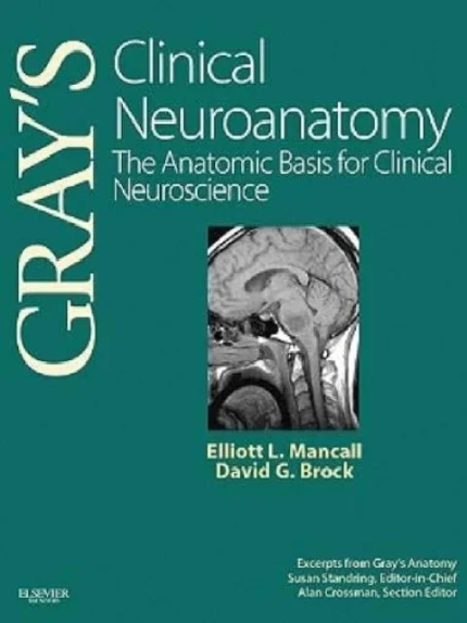
Gray’s Clinical Neuroanatomy: The Anatomic Basis for Clinical Neuroscience (Gray’s Anatomy) 1st Edition

Price range: ₨ 750 through ₨ 1,550
Black & White Print
Color Matt Paper Print
Clear
Gray’s Clinical Neuroanatomy: The Anatomic Basis for Clinical Neuroscience (Gray’s Anatomy) 1st Edition the booklanceby Elliott L. Mancall MD (Author), David G. Brock MD CIP (Author)Gray’s Clinical Neuroanatomy focuses on how knowing functional neuroanatomy is essential for a solid neurologic background for patient care in neurology. Elliot Mancall, David Brock, Susan Standring and Alan Crossman present the authoritative guidance of Gray’s Anatomy along with 100 clinical cases to highlight the relevance of anatomical knowledge in this body area and illustrate the principles of localization.Master complex, detailed, and difficult areas of anatomy with confidence. View illustrations from Gray’s Anatomy and radiographs that depict this body area in thorough anatomical detail. Apply the principles of localization thanks to 100 brief case studies that highlight key clinical conditions. Tap into the anatomical authority of Gray’s Anatomy for high quality information from a name you trust. Presents the guidance and expertise of a high profile team of authors and top clinical and academic contributors.

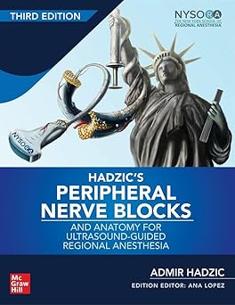
Hadzic’s Peripheral Nerve Blocks and Anatomy for Ultrasound-Guided Regional Anesthesia

Price range: ₨ 1,520 through ₨ 2,620
Black & White Print
Color Matt Paper Print
Clear

Hadzic’s Peripheral Nerve Blocks and Anatomy for Ultrasound-Guided Regional Anesthesia

Hadzic’s Peripheral Nerve Blocks and Anatomy for Ultrasound-Guided Regional Anesthesia is a definitive, clinician-approved reference that bridges anatomy, ultrasound imaging, and regional anesthesia practice. Recognized globally as a cornerstone guide for anesthesiologists, pain specialists, nurse anesthetists, and trainees, this book combines detailed anatomical insight with practical, ultrasound-guided procedural direction — making it essential for both exam preparation and real-world clinical use.

This comprehensive text focuses on ultrasound-guided peripheral nerve blocks, a cornerstone of modern regional anesthesia that enhances precision, safety, and patient outcomes. What sets Hadzic’s book apart is its clear integration of functional anatomy with sonoanatomy — presenting anatomical detail alongside high-quality ultrasound images and procedural illustrations so you understand what you are targeting and why it matters. Every block is explained step-by-step, with emphasis on probe positioning, needle trajectory, safety considerations, and clinical pearls that experienced practitioners rely on.

📘 Key Features

  • 🧠 Comprehensive and Authoritative: Trusted reference for regional anesthesia and ultrasound-guided blocks

  • 📚 Anatomy + Sonoanatomy Focus: Integrates detailed anatomical descriptions with corresponding ultrasound views

  • 🖼️ High-Quality Ultrasound Images & Diagrams: Visual guidance to improve pattern recognition and procedural accuracy

  • 🩺 Step-by-Step Techniques: Systematic approach for performing a wide range of peripheral nerve blocks

  • 💡 Clinical Pearls & Safety Tips: Insights that refine technique and optimize patient safety

  • 🎓 Exam & Clinical Practice Ready: Ideal for exam preparation, training courses, and daily clinical use

  • 🚚 Fast Delivery Across Pakistan: Delivered within just 3 days
McMinn’s Concise Human Anatomy 2nd Edition

Price range: ₨ 500 through ₨ 1,050
Black & White Print
Color Matt Paper Print
Clear
McMinn’s Concise Human Anatomy 2nd Edition the booklanceby David Heylings (Author), Stephen W. Carmichael (Author), Samuel John Leinster (Author), Janak Saada (Author)Focusing on the essentials, McMinn’s Concise Human Anatomy is a convenient, portable guide and revision aid. The clear, jargon-free text is supported by high-quality, labelled photographs of cadaver dissections and surface anatomy, radiological images captured using the latest technologies and explanatory line diagrams, all redrawn for this edition. Providing full explanations of difficult anatomical relationships, and highlighting features of clinical significance throughout, this second edition remains an invaluable guide for students of anatomy across the medical and health sciences, and a handy reference for the busy clinician.

MCQ Companion to Applied Radiological Anatomy 1st Edition

Original price was: ₨ 500.Current price is: ₨ 380.

MCQ Companion to Applied Radiological Anatomy 1st Edition

MCQ Companion to Applied Radiological Anatomy 1st Edition is a targeted resource designed for medical students, radiology trainees, and postgraduate candidates seeking to master applied radiological anatomy through exam-style practice. Authored by experienced radiologists, this book complements traditional anatomy and radiology textbooks by providing a focused, question-based approach that enhances understanding and retention.

This first edition contains a wide collection of multiple-choice questions (MCQs) specifically aligned with applied radiology, covering key areas such as neuroimaging, thoracic imaging, abdominal imaging, musculoskeletal radiology, and head & neck anatomy. Each question is carefully crafted to test both factual knowledge and clinical reasoning, simulating real exam conditions.

Key Features:

  • ✔️ Extensive MCQ collection covering all applied radiological anatomy topics
  • ✔️ Detailed explanations for each question to reinforce learning
  • ✔️ Chapter-wise organization for systematic revision
  • ✔️ Focuses on both factual knowledge and clinical reasoning
  • ✔️ Ideal for FRCR, FCPS, USMLE, and other radiology exams
  • ✔️ Enhances image interpretation and exam readiness
  • ✔️ Perfect for self-assessment and last-minute revision
  • ✔️ Portable and easy-to-use format for quick study
Master applied radiological anatomy and improve your exam performance with MCQ Companion to Applied Radiological Anatomy 1st Edition. Order now from TheBookLance and receive it anywhere in Pakistan within just 3 days with COD service!

Netter’s Atlas of Anatomy for Speech, Swallowing, and Hearing 2nd Edition

Price range: ₨ 500 through ₨ 1,000
Black & White Print
Color Matt Paper Print
Clear
Netter’s Atlas of Anatomy for Speech, Swallowing, and Hearing 2nd Edition the booklanceby David H. McFarland (Author)Filled with Dr. Frank Netter’s world-class illustrations and all the essential information on anatomy and physiology relevant to SLH, Netter’s Atlas of Anatomy for Speech, Swallowing, and Hearing, 2nd Edition uses a unique “read-it, see-it” approach to help you easily connect anatomy and physiology concepts to detailed illustrations. This full-color SLH-specific atlas contains a basic overview of anatomical organization systems, expanded content on swallowing, and updated discussions on the anatomical and functional bases of normal speech, swallowing, and hearing to give you a solid foundation in learning how to diagnose and treat SLH disorders.UNIQUE! Frank Netter’s medical illustrations and straightforward descriptions clearly present the anatomy and physiology of speech, language, and hearing to help you easily build the foundation necessary to diagnose and treat disorders. System musculature summary tables for each section present vital information in a quick, easy, and consistent format for study and reference. Evolve student resources include video clips of cadaver dissections, animations, self-test questions and exercises to enhance your understanding of SLH anatomy and physiology. A balance of text and illustrations with text on the left hand page and the related image on the right provides both the visual and written information you need to know in a “read-it, see-it” format. • NEW! Expanded content on swallowing meets the growing need for this vital information.• NEW! Updated content and references gives you the most current information on the anatomy and physiology relevant to speech, language, and hearing.

Normal Ultrasound Anatomy of the Musculoskeletal System A Practical Guide

Price range: ₨ 300 through ₨ 500
Black & White Print
Color Matt Paper Print
Clear
Normal Ultrasound Anatomy of the Musculoskeletal System: A Practical Guide the booklanceby Enzo Silvestri (Author), Alessandro Muda (Author), Luca Maria Sconfienza (Author)The book provides a comprehensive description of the ultrasound anatomy of the musculoskeletal system and clear guidance on the technique. Ultrasound images are coupled with anatomic pictures explaining probe positioning and scanning technique for the various joints of the musculoskeletal system: shoulder, elbow, hand and wrist, hip, knee, foot, and ankle. For each joint there is also a brief explanation of normal anatomy as well as a list of tricks and tips and advice on how to perform the ultrasound scan in clinical practice. This book will be an excellent practical teaching guide for beginners and a useful reference for more experienced sonographers.

Principles of Anatomy & Physiology 16th Edition

Original price was: ₨ 5,500.Current price is: ₨ 5,300.

Principles of Anatomy & Physiology 16th Edition

Principles of Anatomy & Physiology 16th Edition is a globally trusted textbook that provides a comprehensive understanding of the structure and function of the human body. Designed for medical, nursing, pharmacy, physiotherapy, and allied health students, this updated edition presents complex anatomical and physiological concepts through clear explanations, detailed full-color illustrations, clinical applications, and interactive learning features. It covers all major body systems, homeostasis, cellular physiology, tissues, organs, and the integration of body functions while incorporating the latest scientific research and healthcare advancements. Chapter summaries, review questions, clinical case studies, and learning objectives help reinforce key concepts and improve examination performance. Whether you are preparing for university exams or professional healthcare practice, this textbook is an essential learning resource. Thebooklance offers Principles of Anatomy & Physiology 16th Edition at a discounted price with Cash on Delivery (COD) all over Pakistan within 3–4 working days. Order your copy today.
Rapid Review Gross and Developmental Anatomy

Price range: ₨ 500 through ₨ 1,100
Black & White Print
Color Matt Paper Print
Clear
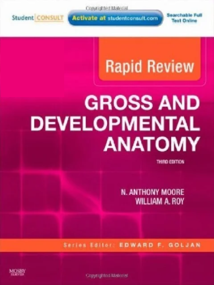
Rapid Review Gross and Developmental Anatomy the booklanceAuthor(s):N. Anthony Moore PhD, William A. Roy PT PhDGet the most from your study time, and experience a realistic USMLE simulation with Rapid Review Gross and Developmental Anatomy, 3rd Edition, by Drs. N. Anthony Moore and William A. Roy. This new reference in the highly rated Rapid Review Series is formatted as a bulleted outline with photographs, tables and figures that address all the gross and developmental anatomy information you need to know for the USMLE. And with Student Consult functionality, you can become familiar with the look and feel of the actual exam by taking a timed or a practice test online that includes 350 USMLE-style questions. Review the most current information with completely updated chapters, images, and questions. Access all the information you need to know quickly and easily with a user-friendly, four-color outline format that includes High-Yield Margin Notes. Take a timed or a practice test online with more than 350 USMLE-style questions and full rationales for why every possible answer is right or wrong. Profit from the guidance of series editor, Dr. Edward Goljan, a well-known author of medical study references, who is personally involved in content review. Get a better understanding of complex anatomical concepts with additional radiologic images as well as anatomical illustrations by Dr. Frank H. Netter. Study and take notes more easily with the new, larger page size. Practice with a new testing platform on USMLE Consult that gives you a realistic review experience and fully prepares you for the exam.

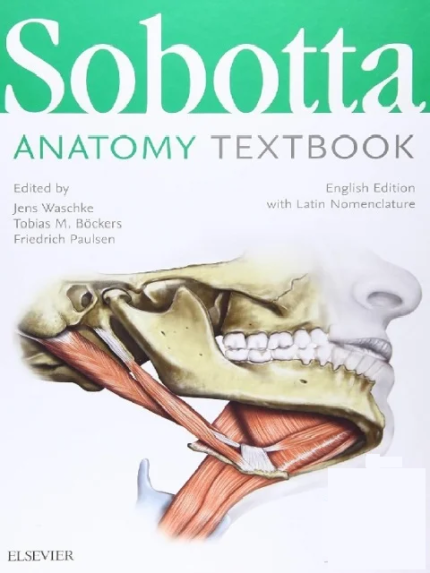
Sobotta Anatomy Textbook: English Edition with Latin Nomenclature 1st Edition

Price range: ₨ 1,450 through ₨ 2,950
Black & White Print
Color Matt Paper Print
Clear
Sobotta Anatomy Textbook: English Edition with Latin Nomenclature 1st Edition the booklanceby Friedrich Paulsen (Editor), Tobias M. Böckers (Editor), Jens Waschke (Editor), Stephan Winkler (Illustrator), Katja Dalkowski (Illustrator), Jörg Mair (Illustrator), Sonja Klebe (Illustrator)Like the Sobotta Atlas of Anatomy, this textbook guides students through the anatomy in a clear, structured manner that is easy to remember. Sobotta excels at comprehensibly explaining the fascinating world of anatomy and carries you safely through every test and your initial patient interactions.Clinical Cases: Presents a relevant practical medical case as it will be encountered during rotation . It highlights how students can gain valuable insights into the case by taking structured notes during patient examination.Clinical Remarks: Indicate functional and clinical aspects that help to identify a topic’s relevance and put it in context for subsequent patient diagnosis and treatmentSkills Boxes: Provide a quick overview of competency-based study objectives, which help you build a contextualised knowledge useful in exams and applicable to future professional practice.Regular Note Boxes: Highlight particularly important exam knowledge to be retained throughout the bookThe textbook fosters integrated learning by linking macroscopic anatomy, where relevant, to embryology. The detailed chapter on the anatomy of the head makes this textbook particularly suitable for students of dental medicine.The Sobotta Anatomy Textbook enables you to develop a true contextualised understanding of the subject and thus not just pass anatomy exams, but build a foundation for your future clinical work!

Teaching Anatomy: A Practical Guide 2nd ed

Price range: ₨ 1,000 through ₨ 2,050
Black & White Print
Color Matt Paper Print
Clear
Teaching Anatomy: A Practical Guide 2nd ed the booklanceby Lap Ki Chan (Editor), Wojciech Pawlina (Editor)The field of anatomy is dynamic and fertile. The rapid advances in technology in the past few years have produced exciting opportunities in the teaching of gross anatomy such as 3D printing, virtual reality, augmented reality, digital anatomy models, portable ultrasound, and more. Pedagogical innovations such as gamification and the flipped classroom, among others, have also been developed and implemented. As a result, preparing anatomy teachers in the use of these new teaching tools and methods is very timely.The main aim of the second edition of Teaching Anatomy – A Practical Guide is to offer gross anatomy teachers the most up-to-date advice and guidance for anatomy teaching, utilizing pedagogical and technological innovations at the forefront of anatomy education in the five years since the publication of the first edition.This edition is structured according to the teaching and learning situations that gross anatomy teachers will findthemselves in: large group setting, small group setting, gross anatomy laboratory, writing examination questions, designing anatomy curriculum, using anatomy teaching tools, or building up their scholarship of teaching and learning. Fully revised and updated, including fifteen new chapters discussing the latest advances, this second edition is an excellent resource for all instructors in gross anatomy.

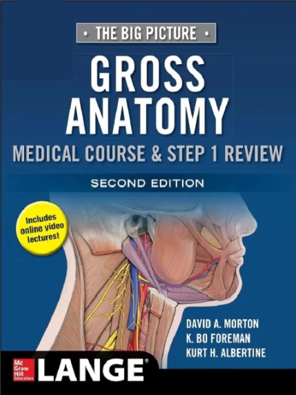
The Big Picture Gross Anatomy Medical Course & Step 1 Review 2nd Ed

Price range: ₨ 850 through ₨ 1,750
Black & White Print
Color Matt Paper Print
Clear
The Big Picture Gross Anatomy Medical Course & Step 1 Review 2nd Ed the booklanceby David Morton (Author), K. Bo Foreman (Author), Kurt Albertine (Author)The Big Picture: Gross Anatomy, Second Edition is the perfect bridge between reviews and textbooks. With an emphasis on what you truly need to know versus “what’s nice to know”, it features more than 400 full-color illustrations, along with a simple, effective page design that gives you a complete, yet concise, overview of essential anatomy.The book’s user-friendly presentation consists of text on the left-hand page and beautiful full-color illustrations on the right-hand page. In this way, you get a “big picture” of anatomy principles, delivered one concept at a time – making them easier to understand and remember.Zero-in on what you really need to know to ace the course and board exams with: NEW! Access to high-quality videos on each major topic — students can view them before reading the text or attending lectures Key structures highlighted in bold when they are first mentioned Bullets and numbers used to break down important concepts More than 400 full-color figures illustrate essential anatomy High-yield clinically relevant concepts indicated by a special icon Study questions and answers following each section A final exam at the end of the book If you want a well-illustrated, succinct, high-yield review to help you excel on your coursework and the boards, your search ends with The Big Picture: Gross Anatomy, Second Edition.

The Big Picture Gross Anatomy Medical Course & Step 1 Review 2nd Ed (Copy)

Price range: ₨ 500 through ₨ 1,100
Black & White Print
Color Matt Paper Print
Clear
A Radiologically-Guided Approach to Musculoskeletal Anatomy the booklanceby Alberto Tagliafico (Author), Carlo Martinoli (Author)For many healthcare professionals, musculoskeletal diseases represent the “bread and butter” topic after graduation. Therefore, radiological education in respect of the musculoskeletal system is vital in ensuring adequate patient management and cost-effective use of healthcare financial resources. This book illustrates the clinical anatomy of the musculoskeletal system by means of images obtained using commercially available imaging equipment and the three main imaging techniques employed today – magnetic resonance imaging, computed tomography, and ultrasound. Based on an integrated multimodality approach, each anatomical region is presented with a special focus on clinically relevant anatomical details and the characteristic findings observed in patients referred by physicians. With almost 450 images and illustrations, A Radiologically Guided Approach to Musculoskeletal Anatomy is intended as a bridge from a standard anatomical atlas to diagnostic imaging. It will assist in the everyday interpretation of imaging studies of the musculoskeletal system, providing prompt answers to frequently encountered questions. Clinical notes and self-assessment modules are also provided. All who wish to learn more about the role of diagnostic imaging of the musculoskeletal system will find this book to be of great value. It will benefit not only medical students and residents but also radiology technologists and professionals in other fields of health care, including orthopaedists, rheumatologists, and rehabilitation specialists. ​

Ultrasonic Topographical and Pathotopographical Anatomy

Price range: ₨ 350 through ₨ 650
Black & White Print
Color Matt Paper Print
Clear
Ultrasonic Topographical and Pathotopographical Anatomy the booklanceby Z. M. Seagal (Author), O. V. Surnina (Author)Written by experienced and well-respected physicians and professors, this new all-color volume presents the ultrasonic topographical and pathotopographical anatomy of the body, including the head, neck, chest, anterolateral abdominal wall, abdominal organs, retroperitoneal space, male and female pelvises, and lower extremities.Specific and non-specific ultrasonic symptoms are suggested for normal and abnormal developmental variants, diffuse and local pathotopographical anatomy. This color atlas contains comparative topographical and pathotopographical data and is the first manual of its kind for students and medical specialists in different areas, including those specializing in medical sonography. The original technology was tested at clinics in patients subjected to ultrasonic monitoring. Because of early detection there were no false-positive or false-negative results. The therapy was effective, and, in some cases, the use of the original method of “seagalography” (optometry and pulsemotorgraphy) has made it possible to develop new methods of treatment and/or to determine the optimal doses of drugs, as well as to develop effective drug complexes for treatment of a given pathology.This important new volume will be valuable to physicians, junior physicians, medical residents, lecturers in medicine, and medical students alike, either as a textbook or as a reference. It is a must-have for any physician’s library.